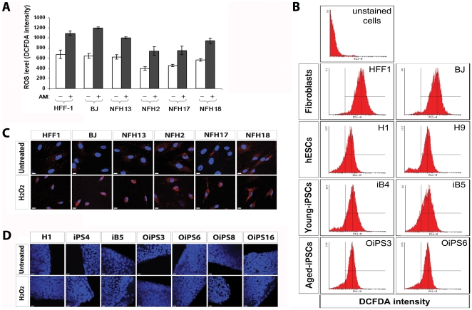
Figure 7. ROS and DNA damage in aged donor-derived iPSCs.
(A) Production of ROS in fibroblast cells was measured by FACS using DCF-DA at basal level and after 24 h treatment with high doses actinomycin D (AM) (10−6 M). Analysis was conducted in quadruplicate and reported as fluorescence intensity. Error bars indicate standard deviation. (B) Representative DCF-DA-based FACS measurements of basal ROS generation in two fibroblasts (HFF1 and BJ), two hESC lines (H1 and H9), two young donor-derived iPSC lines (iB4 and iB5), and two aged donor-derived iPSC lines (OiPS3 and OiPS6). (C) Oxidative damage to nuclear and mitochondrial DNA within fibroblast cells was assessed by 8-hydroxy-2′-deoxyguanosine (8OHdG) staining. Treatment with 500 µM H2O2 for two hours was employed to trigger oxidative-mediated DNA damage. Scale bars, 10 µm. (D) Embryonic and somatic-derived pluripotent stem cells were stained with an antibody against 8OHdG to detect nuclear or mitochondrial DNA lesions induced by oxidative stress. The analysis was conducted in standard growth conditions and after a 2 h exposure to 500 µM H2O2. Scale bars, 10 µm.

