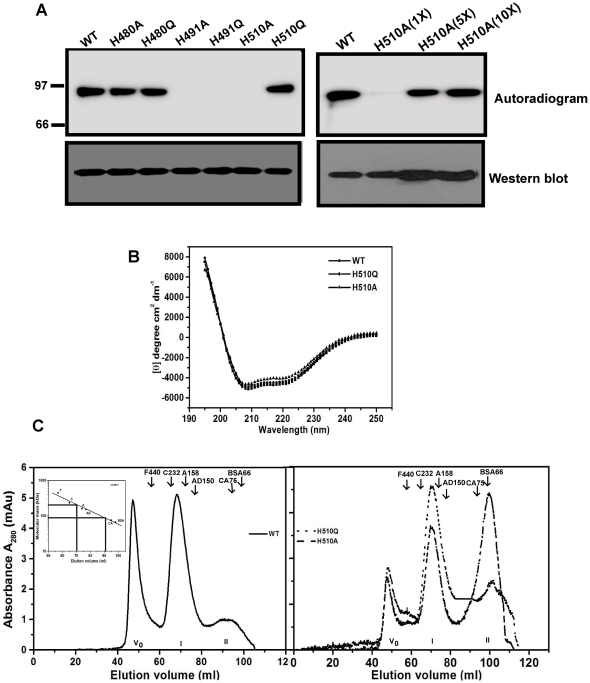
Figure 1. mPPK1 autophosphorylates at His-491 residue.
A. Autophosphorylating ability of mPPK1 in response to mutations at conserved histidine residues. Upper left panel: Autophosphorylation activities of wild-type (lane 1) and mutant proteins (lanes 2–7) using 500 ng protein/reaction. Upper right panel: Autophosphorylation of H510A mutant protein with increasing concentrations (1X = 500 ng; 5X = 2.5 µg and 10X = 5 µg) of protein. Western blotting of mutant proteins was carried out using anti-His antibody (lower left and right panels). B. Far-UV CD spectra of mutant proteins. C. Gel filtration elution profile of wild-type (WT) and mutant (H510A and H510Q) proteins. Vo indicates void volume. I and II represent the molecular mass corresponding to dimeric and monomeric forms of mPPK1 respectively. Inset: Molecular mass calibration curve using different proteins. Notation used: F, Ferritin (440 kDa); C, Catalase (232 kDa); A, Aldolase (158 kDa); AD, Alcohol dehydrogenase (150 kDa); CA, Conalbumin (75 kDa) and BSA, Bovine serum albumin (66 kDa). Position of molecular mass markers is indicated.

