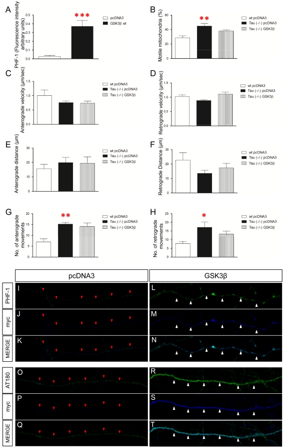
Figure 5. GSK3β overexpression effects are mediated by Tau phosphorylation.
A: Tau phosphorylation is increased after GSK3β overexpression (F1,18 = 20.987; p = 0.0001). B: Percentage of motile mitochondria in Tau (−/−) neurons is increased compared to wt (F1,26 = 9.34; p = 0.001) whereas GSK3β overexpression in Tau (−/−) neurons does not modify this parameter. C–D: Anterograde (C) or retrograde (D) mitochondrial velocity in the absence of tau and after overexpression of GSK3β. E–H: Tau absence does not modify mitochondria run length but increases the number of anterograde and retrograde movements. E–F: Neither anterograde (E) nor retrograde (F) run length is modified in the absence of Tau or after overexpression of GSK3β. G–H: Number of anterograde (G) and retrograde (H) movements in the absence of tau and after overexpression of GSK3β in Tau (−/−) neurons. Tau absence increases the number of both anterograde (F1,26 = 9.164; p = 0.001) and retrograde movements (F1,28 = 4.992; p = 0.015) compared to wt neurons. I–T: Phosphorylation of tau is increased after GSK3β transfection. PHF-1 staining in GSK3β- (L–N) and pcDNA3-transfected axons (I–K). PHF-1: Green; myc: Blue. Similarly, AT180 staining is also increased in GSK3β- (R–T) compared to pcDNA3-transfected axons (O–Q). AT180: Green; myc: Blue. Red triangles indicate pcDNA3-transfected axons. White triangles label GSK3β-transfected axons. Red asterisk: * 0.05>p>0.01; ** 0.01>p≥0.001; *** 0.001>p≥0.0001.

