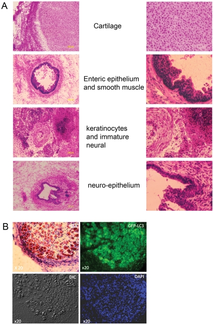
Figure 2. Teratoma sections derived from HES3 LC3-GFP cells and persistence of LC3-GFP transgene expression.
(A), Representative sections of a 6 week old teratoma derived from HES3-LC3-GFP cells showed cell types representative of the three germlayers with each section shown magnified at right. (B), GFP fluorescence persists in teratomas of HES3-LC3-GFP cells. Representative images showing haemtoxylin/eosin staining and GFP fluorescence of two immediately adjacent serial sections of a HES3-LC3-GFP teratoma are shown. Brightfield and DAPI staining images corresponding to the GFP-fluorescence panel are shown. Arrow heads highlight autophagosomes in themagnified section (inset).

