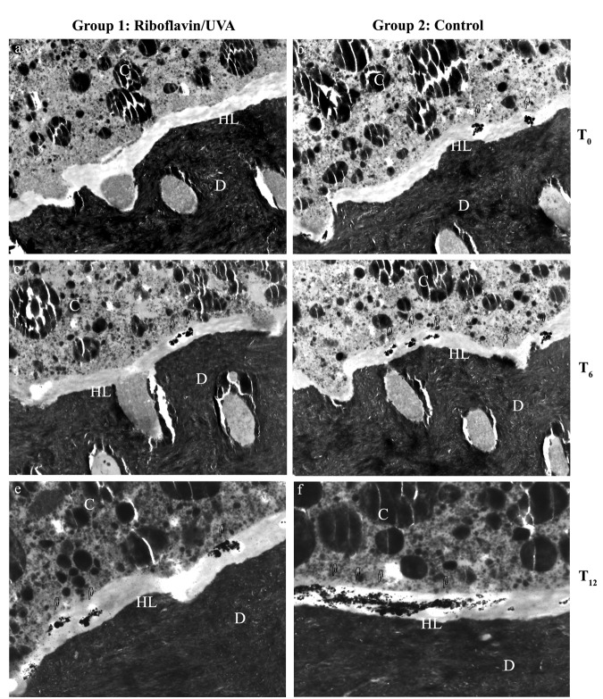
Figure 2.
Transmission electron microscopy images showing interfacial nanoleakage expression. D, dentin; HL, hybrid layer; C, composite resin. Original magnification: 4600×. Adhesive interfaces created by XP Bond after riboflavin/UVA treatment (Figs. 2a, 2c, 2e) or on control (Figs. 2b, 2d, 2f), respectively. Scattered deposits of silver nitrate were found immediately after bonding on riboflavin/UVA-treated specimens (T0, Fig. 2a) and on control specimens (T0, Fig. 2b). After 6 mos of storage in artificial saliva at 37°C, both the riboflavin/UVA-treated interface (T6, Fig. 2c) and the control specimens (T6, Fig. 2d) showed similar silver deposits with increased nanoleakage compared with T0. After 12 mos of storage, silver deposits were recorded in the riboflavin/UVA-treated group (T12, Fig. 2e), and large areas of silver diffused throughout the hybrid layer were frequently found in controls (T12, Fig. 2f).

