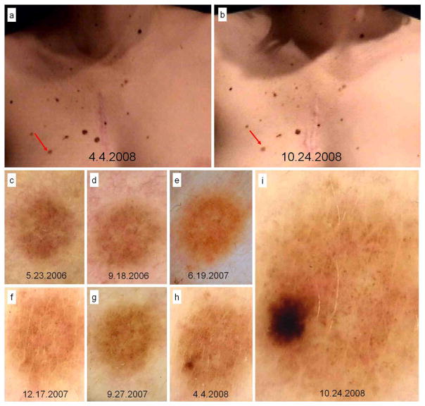
Figure 2.
In situ melanoma developed over melanocytic nevus in a 23 year-old patient, with personal and familial history of melanoma, diagnosed due to changes in digital follow-up. Body-mapping images displaying no clinical change (A and B) and dermoscopy records in chronological order until excision after 29 months and 7 visits of follow-up (C to I).

