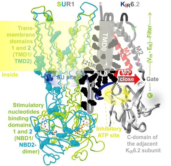
Fig. 1.
A working model of a KATP gating unit. One quarter of KATP complex is shown for clarity. ND ABCC8 mutations are mapped as balls. The SUR1 core is shown in a backbone wire presentation. Nucleotides are rendered in licorice style. KIR6.2 pore-forming domains are presented as ribbons. No high resolution structural template is available for the N-terminal domain of SUR1 or the KIR N-tail. L213R and other elements of the model discussed in the text are identified using color-coded labels. The model of SUR1/KIR6.2 coupling predicts the KIR-closing movement of L0 and M0 helices colocalized at the membrane-cytosol interface.

