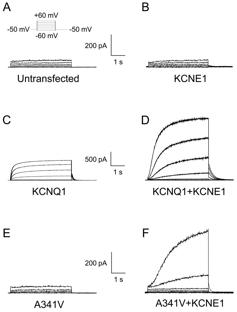
Fig. 1.
Representative whole cell current traces recorded from untransfected and transiently transfected HL-1 cardiomyocytes. A. Current recording from an untransfected HL-1 cardiomyocyte is shown. A small, time-independent background current was detectable. The inset shows the 4-s test pulse voltage protocol. B. Current recording from an HL-1 cell transiently expressing KCNE1 β-subunit is presented. No difference was observed compared to the untransfected HL-1 cardiomyocyte. C. HL-1 cell expressing the KCNQ1 α-subunit resulted in functional channels with fast activation kinetics. D. The co-expression of KCNQ1 with KCNE1 resulted in functional channels with slow activation kinetics, characteristic of native wild type IKs. E. The KCNQ1 A341V mutant did not result in functional channels. No differences were observed compared to the untransfected cells. F. The co-expression of A341V with KCNE1 resulted in functional channels with markedly slow activation kinetics.

