Fig. 3.

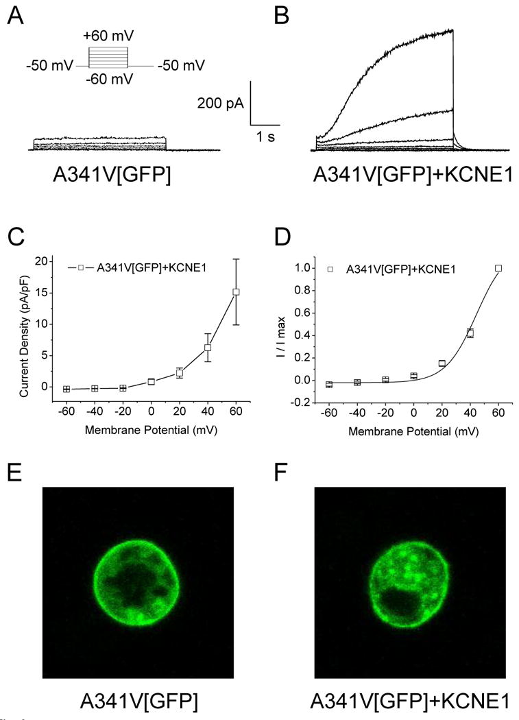
Cellular localization of A341V expressed in HL-1 cells. A GFP-tagged A341V (A341V[GFP]) construct was transiently expressed in HL-1 cells. Representative current traces of A341V[GFP] in the absence (A) or co-expressed with KCNE1 (B) are shown. The current-voltage relationship, normalized to cell capacitance (C) and the isochronal activation curve (D) for A341V[GFP]+KCNE1 (n=7) revealed no significant changes compared to those of A341V+KCNE1 (in the absence of GFP; Table 1). Cellular localization of A341V[GFP] in the absence (E) or co-expressed with KCNE1 (F) were visualized by confocal microscopy. In both cases, surface expression of the mutant protein was evident.
