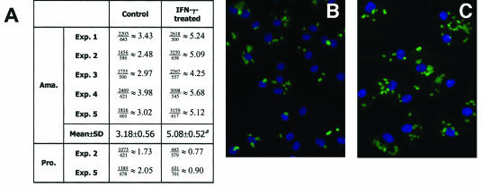
FIG. 2.
Microscopic evaluation of L. amazonensis infection in MΦs. BM-MΦs from BALB/c mice (Exp. 1 and 2) or C57BL/6 mice (Exp. 3, 4, and 5) were seeded at a concentration of 1.5 × 10⁵ cells/chamber on chamber slides. Cells were not treated or were treated with 20 ng of IFN-γ per ml for 4 h prior to infection with 3 × 10⁵ amastigotes (Ama.) or 7.5 × 10⁵ stationary-phase promastigotes (Pro.). After 48 h of incubation, cells were processed for immunostaining of the parasites. (A) Summary of the average numbers of parasites per cell, as calculated by dividing the total number of parasites by the total number of MΦs examined. (B and C) Representative images of amastigote-infected control MΦs (B) and IFN-γ-treated MΦs (C). A superscript a indicates that the P value is <0.01.

