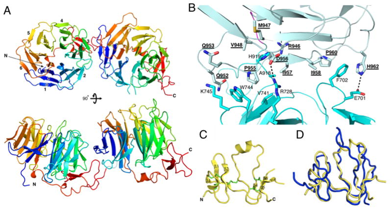
Figure 2. Structures of LRP6(3-4) and Dkk1_C.
(A) The LRP6(3-4) structure, viewed down the pseudo-six fold symmetry axis of propeller 3 (top) or towards the side (bottom). Blades are numbered in repeat 3, and individual strands are labeled in blade 1.
(B) Closeup of the repeat 3-4 interface. Interacting side chains are shown in stick representation. Polar interactions are shown with dashed lines. Repeat 4 residue labels are underlined. The complete list of interactions is given in Table S2. See also Figure S1.
(C) Ribbon representation of Dkk1_C. Disulfide bridges are show in green. The loop between β-strands 5 and 6 is disordered. See also Figure S2
(D) Superposition of human Dkk1_C structure (gold) with the NMR solution structure of mouse Dkk2_C (blue) (PDB entry 2JTK).

