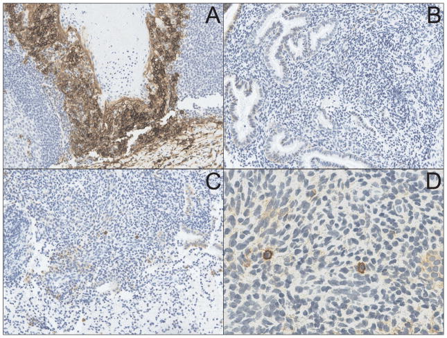
Fig. 1.
CD138 immunostaining is superior to MGP staining of endometrial biopsy sections for identification of plasma cells. (A) Immunohistochemical staining of tonsilar tissue (used as positive controls) with the plasma cell-specific marker CD138 (syndecan-1) shows characteristic membrane staining (X200). (B) Endometrial stroma from a woman originally diagnosed with chronic endometritis (upon identification of plasma cells with MGP staining) demonstrates complete absence of CD138-positive cells (X200). (C) Endometrial biopsy sample from a woman originally diagnosed with normal endometrial tissue shows multiple CD138-positive cells in the endometrial stroma (X200) (D). CD138 positive endometrial cells from section show in Fig. 1C display cell membrane staining characteristically similar to that seen in tonsilar tissue (X600).

