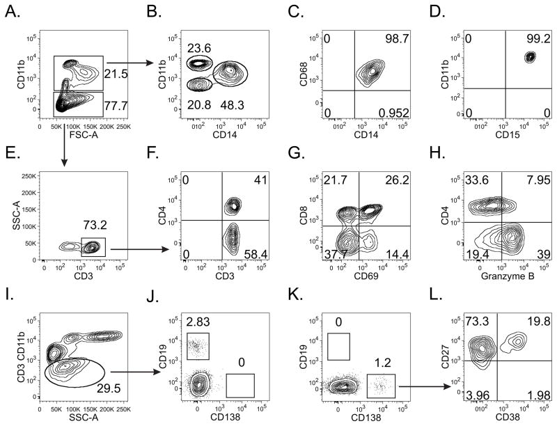
Fig. 3.
Flow cytometric analysis of endometrial biopsies represents a powerful approach for identification of upper genital tract leukocytes (representative contour plots shown). After defining the CD45+ cell population (not shown), a large percentage was identified as (A) CD11b+, and (B, C) further identified as macrophages (CD11int/hiCD14+CD68+) (B, D) neutrophils (CD11bhiCD14-CD15+), and (B) another myeloid cell subpopulation (CD11bloCD14−CD11−). (E) To characterize T cell populations, CD11b− cells were interrogated for CD3 expression. Both CD4+ and CD8+ cell populations were clearly defined (F), allowing exploration of their functional activation status, as indicated by the levels of CD69 (G) and granzyme B (H) expression. For plasma cell identification, CD3 and CD11b− cells were interrogated for CD19 (J) and CD138 (K) expression. All CD138+ cells were CD27+ while a significant portion were CD38+ (L), strong evidence for the proper identification of a plasma cell population.

