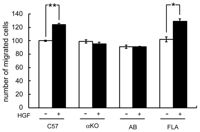
Fig 4. Myoblasts from the α-syntrophin-knockout mice do not respond to HGF stimulation.
Skeletal myoblasts were isolated from C57, α-syntrophin-knockout (αKO), α/β2-syntrophin-double knockout (AB), and α-syntrophin transgenic (FLA) mice and seeded on the insert of a three dimensional chamber assay as described in “Materials and Methods”. After incubation for 1 h, cells were fixed with cold methanol and stained with hematoxylin. The cells migrating to the lower chamber were counted using an inverted microscope. Values are expressed as means ± S.E.M. from four independent experiments (*p<0.01, **p<0.001).

