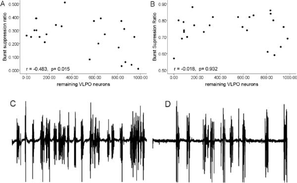
Figure 3.
The relationship between the number of VLPO neurons and the depth of anesthesia.
Top: Correlation between the number of remaining VLPO neurons following lesions and the burst suppression ratio at steady state isoflurane A) 1% and B) 2%. Bottom: Sample EEG traces (1 min duration) during 1% (C) and 2% (D) isoflurane.

