
Figure 7.
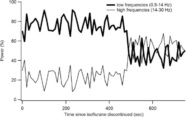
Time course of the return of high frequency EEG power at recovery from anesthesia. Shown is the relative low frequency (0.5–14 Hz) and high frequency (14–30 Hz) EEG power following discontinuation of isoflurane in one rat (R3924). Note the abrupt transition that we took as electrographic emergence (Fig 8).