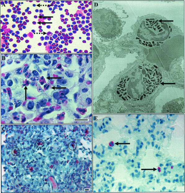
FIG. 1.
Accumulation of eosinophils in experimental pulmonary tuberculosis in the guinea pig. (A) Representative photomicrograph of bronchoalveolar cells collected at 5 weeks postinfection with M. tuberculosis, with eosinophils (solid black arrows), large mononuclear cells (right-pointing dashed arrow), and lymphocytes (left-pointing dashed arrow). Magnification, ×40. (B) Representative photomicrograph of a guinea pig tuberculous granuloma 11 days after an aerosol infection. Note the multiple eosinophils (black arrows). Hematoxylin and eosin stain was used. Bar = 10 μm. (C) Representative photomicrograph of a guinea pig tuberculous granuloma 11 days after aerosol infection, stained with Luna's eosinophil granule stain. Positive cells are surrounded by black rings. Bar = 10 μm. (D) Representative transmission electron micrograph of a guinea pig tuberculous granuloma 11 days after aerosol infection. Note the two eosinophils with distinctive intracytoplasmic granules (arrows). (E) Representative photomicrograph of a guinea pig tuberculous granuloma stained for major basic protein at 30 days postinfection with low-dose M. tuberculosis. Magnification, ×100.

