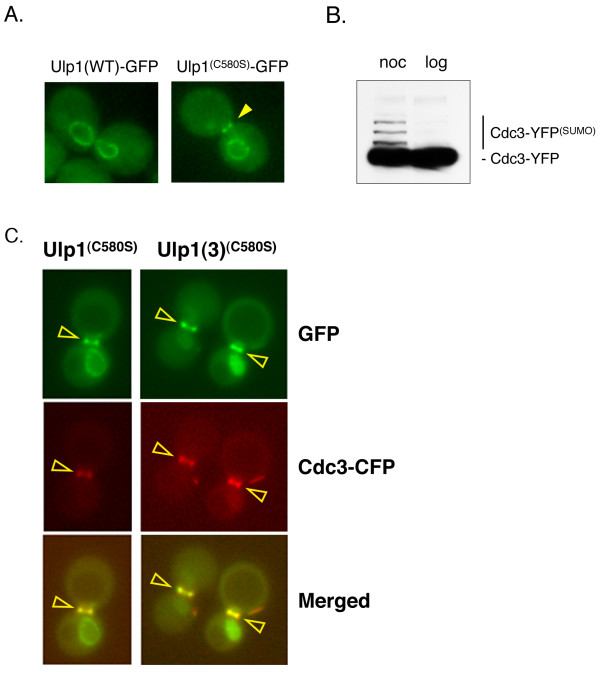
Figure 1.
Localization of Ulp1 and the catalytically inactive Ulp1(C580S) in dividing yeast cells. (A) Yeast cells (MHY500) were transformed either with a low-copy plasmid expressing GFP fusions of Ulp1 or with the catalytically inactive Ulp1(C580S) mutant. Representative images indicating the localization of GFP-tagged Ulp1 and Ulp1(C580S) after nocodazole-induced G2/M arrest are shown (YOK 1611 and YOK 1474). Note that only the Ulp1(C580S) mutant can be seen at the bud-neck of arrested cells. The arrowhead indicates the position of the bud-neck. (B) Confirmation of sumoylation of Cdc3 was achieved. Whole-cell extracts (WCE) from yeast cells expressing the YFP-tagged septin Cdc3 (YOK 1398) were treated with nocodazole (noc) or grown logarithmically (log) prior to preparation of WCEs. Extracted proteins were then separated on SDS-PAGE gels and probed with the JL-8 antibody (see Methods) to detect Cdc3-YFP and more slowly migrating sumoylated Cdc3-YFP adducts. The identity of sumoylated Cdc3-YFP bands was confirmed by comparing gel shift assays with untagged and FLAG-tagged Smt3 (data not shown). (C) Colocalization of Cdc3 and Ulp1 is shown. A strain coexpressing full-length Ulp1(C580S)-GFP (green) and Cdc3-CFP (red) (strain YOK 2204) was arrested in G2/M and then observed under a fluorescence microscope with the appropriate filter sets (left panel). Arrowheads indicate septin-localized, pseudocolored Ulp1-GFP (green), Cdc3-CFP (red) and the merged image (overlay). Also shown for comparison (right panel) is the colocalization of the Ulp1(3)(C580S)-GFP truncation and Cdc3-CFP (strain YOK 2205). Ulp1(3)(C580S)-GFP is described in Figure 4.

