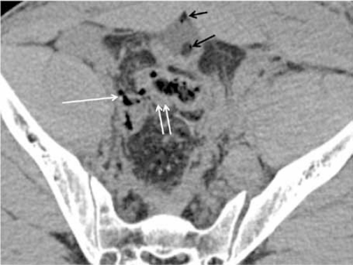
Figure 7a:
CT scan of perforated bowel in a 26-year-old man with MVA. Note subtle extraluminal air (single white arrows) with focal bowel wall thickening (double white arrows) at the rectosigmoid region that was missed on initial review of the CT images. Also note air pockets in the urinary bladder (black arrows). Urinary bladder perforation and transection at the rectosigmoid junction were detected intraoperatively.

