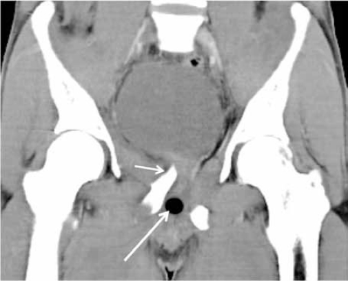
Figure 9b:
CT demonstrating soft tissue injury associated with pelvic fracture. A coronal MPR CT image in soft tissue window of the same patient in Figure 9a showed the fractured fragment (short arrow) compressing at the base of the urinary bladder. Note the mal-positioned Foley’s catheter balloon within the urethra (long arrow). Urethrogram demonstrated a membranous urethral injury.

