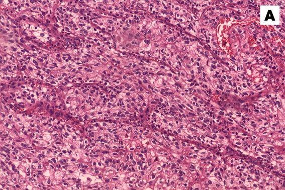
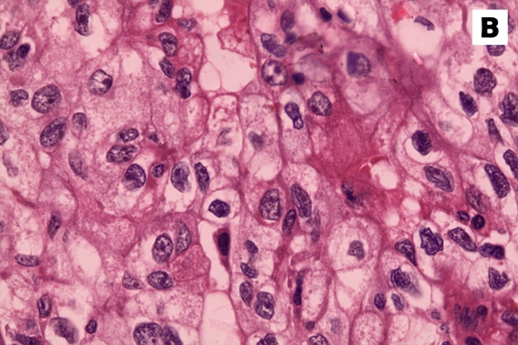
Figure 3:
Haematoxylin and eosin stain of the tumour specimen. (A) A low-power view (10x magnification) showed groups of neoplastic cells separated by fibrous septa with intervening blood vessels. (B) The cell morphology is better appreciated in the high-power view (40x magnification), which clearly revealed the neoplastic features of the cells with pleomorphic nucleus, some with conspicuous nucleoli and abundant, clear to eosinophilic cytoplasm.


