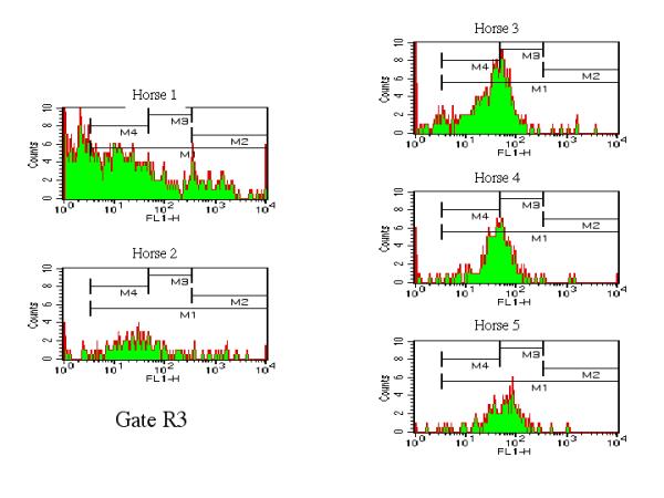
Figure 4.

Flow cytometric representation of morphological and fluorescent 1 (FL 1) signal of platelets. Histogram plots of the fluorescence signal carried by platelets delineated within gate R3. PaIg was determined by performing histogram analysis of the fluorescence carried by the platelets gated into R3. Markers M1, M2, M3 and M4 are identical to the displayed in Figure 2.
