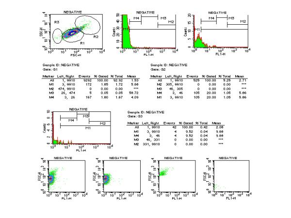
Figure 8.

Protocol for the flow cytometric representation of morphological and fluorescent 1 (FL 1) signal and statistical analysis of platelets before staining (Negative sample) of a healthy Horse. Density plot (FSC × SSC) of unstained and ungated platelets from a normal horse is displayed the upper left row. Histogram plots of the fluorescence signal carried by unstained platelets delineated within gate R1, R2 and R3 are displayed in the upper and middle rows. Marker M1, M2, M3 and M4 are identical to previous figures. Statistical analysis of each gate is displayed in middle rows. The lower row displays the set of four panels (FSC × FL1 plots of platelets). No Gate (external left panel). Gate R1 (internal left panel). Gate R2 (internal right panel). Gate R3 (external right panel)
