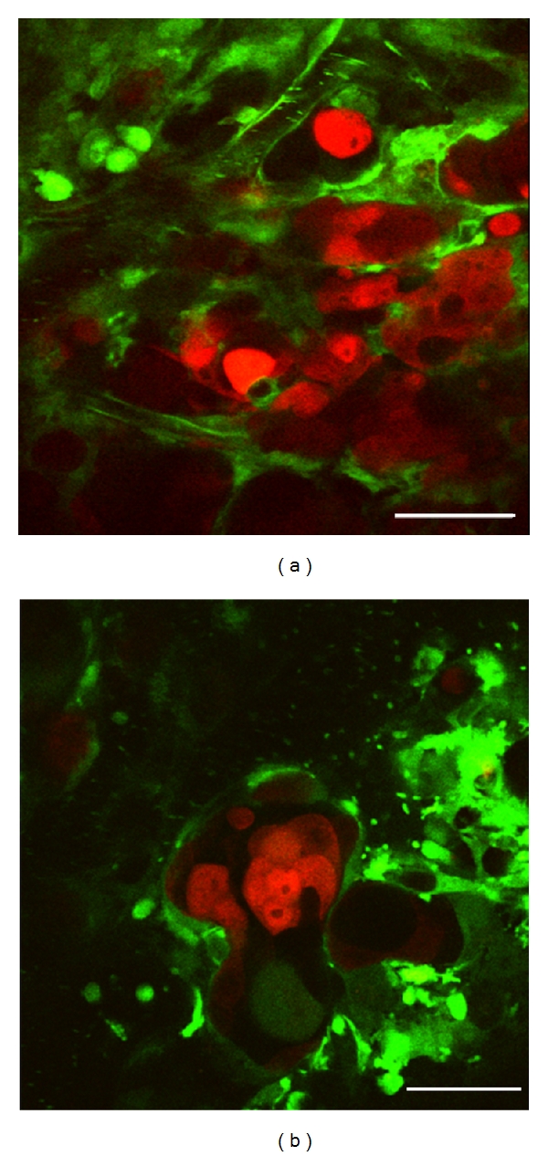
Figure 6.

High-resolution optical imaging of liver metastatic nodule by intravital TPLSM. (a) Liver metastatic nodules were composed of tumor cell clusters and dilated/tortuous vessels (×600, bar, 50 μm). (b) Dilated and tortuous tumor vessels were observed among the clusters of several tumor cells. A flow of aggregated platelets was frequently observed within the tumor vessels (×600, bar, 50 μm).
