
Figure 2.
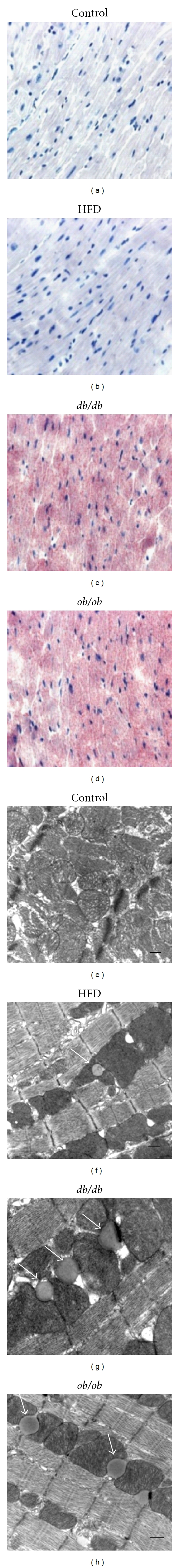
Cardiac histology. ((a), (b), (c), and (d)). Representative Oil-Red-O-(ORO-) stained sections of hearts from (a) control, (b) high fat diet-fed (HFD), (c) db/db, and (d) ob/ob mice. Lipid droplets were rare in control hearts and uncommon in HFD mice despite a ~12-fold increase in cardiac triglyceride content in the latter group. There was an obvious increase in ORO-stainable lipid in the hearts of ob/ob and db/db mice. ((e), (f), (g), and (h)). Transmission electron microscope images of (e) control, (f) HFD, (g) db/db, and (h) ob/ob mice. Lipid droplets (arrows) were rare in control hearts, but individual droplets could be found in many fields in the HFD group. Lipid droplets were larger and much more common in db/db and ob/ob hearts, with multiple droplets typically being observed in most fields. The scale bar indicates 500 nm. Lipid droplets in the heart were almost uniformly in direct contact with mitochondria.