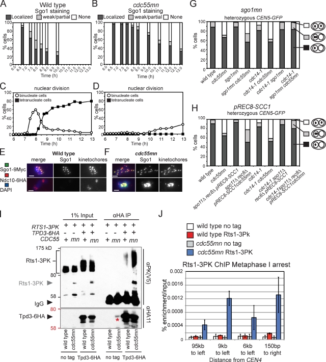
Figure 7.
Overprotection of cohesin in a cdc55mn cell during meiosis. (A–F) Wild-type (AM7228; A, C, and E) and cdc55mn (AM7229; B, D, and F) cells carrying GAL-NDT80 and pGPD1-GAL4(848).ER were released from the pachytene block, and Sgo1 localization at kinetochores (A and B) or the percentages of binucleate and tetranucleate cells (C and D) were determined in a single experiment. (E and F) Examples of Sgo1 localization are shown. Bars, 2 µm. (G and H) Depletion of Sgo1 (G) or replacement of Rec8 by Scc1 (H) abolishes equational segregation in cdc14-1 cdc55mn cells. Strains carrying heterozygous CEN5-GFP and with the indicated genotypes were treated as described in Fig. 1. n = 680–2,000. Strains used were wild type (AM4796), cdc55mn (AM4891), sgo1mn (AM4911), sgo1mn cdc55mn (AM7286), cdc14-1 (AM6910), cdc14-1 cdc55mn (AM6934), cdc14-1 sgo1mn (AM7360), cdc14-1 sgo1mn cdc55mn (AM7421), spo11Δ rec8Δ pREC8-SCC1 (AM5501), spo11Δ rec8Δ pREC8-SCC1 cdc55mn (AM5502), spo11Δ rec8Δ pREC8-SCC1 cdc14-1 (AM7361), and spo11Δ rec8Δ pREC8-SCC1 cdc14-1 cdc55mn (AM7362). (I) Tpd3-6HA was immunoprecipitated using anti-HA antibodies from meiotic extracts of wild-type and cdc55mn cells carrying RTS1-3PK and either TPD3-6HA (AM8028 and AM8014) or no tag (AM8012 and AM8029). Anti-V5 (PK) and anti-HA immunoblots of input and immunoprecipitated samples from strains of the indicated genotypes are shown, with protein molecular mass markers shown in black or red, respectively. An Rts1-3PK degradation product is indicated by the gray arrowhead, and the asterisk indicates residual 3HA-Cdc55. (J) qPCR analysis of chromatin immunoprecipitated using anti-V5 (PK) antibodies from cdc20mn (AM3560), cdc20mn RTS1-3PK (AM7902), cdc20mn cdc55mn (AM7903), and cdc20mn cdc55mn RTS1-3PK (AM7904) strains. The mean of three experiments is shown with error bars indicating standard deviation.

