Figure 1.
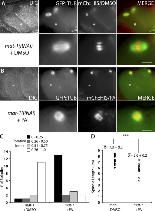
CDK-1 inhibition by PA results in short and rotated meiotic spindles in metaphase-I–arrested APC-depleted meiotic embryos. (A and B) Live images of mat-1(RNAi) meiotic embryos expressing mCherry::histone and GFP::tubulin in utero after treatment with DMSO (A) or PA (B). The bottom panels are magnified images of the top panels. Asterisks mark the +1 embryo. DIC, differential interference contrast. Bars, 5 µm. (C) Quantification of rotation index in mat-1(RNAi) meiotic embryos after treatment with DMSO (left; n = 15) or PA (right; n = 20). The y axis is the number of meiotic embryos. The data shown include measurements from over seven independent injections for each treatment. (D) Quantification of meiotic spindle length in mat-1(RNAi) meiotic embryos after treatment with DMSO (closed circles; n = 15) or PA (open circles; n = 24). Each data point represents an individual meiotic spindle length measurement. The ±SEM is rounded to the nearest tenth. ***, P < 0.0005.

