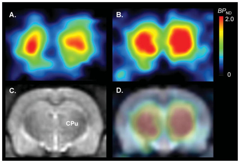
Fig. 2.

Parametric images of [11C]MNPA BPND calculated with a kinetic reference tissue model at baseline (A) and after dopamine depletion (B). Depletion of endogenous dopamine induced by reserpine plus α-methyl-para-tyrosine increased striatal [11C]MNPA BPND by twofold. An image of a T-2 weighted MRI (C) [CPu: caudate putamen (striatum)] and fused PET-MRI image (D) are shown for clearer orientation of striatum.
