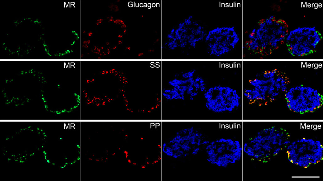
Fig. 5.
The MR is produced in pancreatic delta and pancreatic polypeptide-positive cells in isolated murine islets. Immunofluorescence staining was performed on three consecutive sections of isolated murine islets embedded in collagen (40× objective). The MR (green) localised to the islet periphery and did not co-localise with insulin (blue pseudocolour) or glucagon (red, top row). The MR also co-localised with somatostatin- (SS, red, middle row) and pancreatic polypeptide-positive (PP) cells (red, bottom row). Scale bar 100 µm

