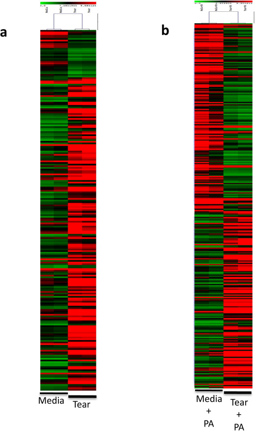
Figure 3. Microarray Analysis of Epithelial Cells Exposed to Tear and/or P. aeruginosa antigens.
a, b Heat-maps of differentially regulated probe sets in epithelial cells incubated with human tear fluid for 6 h (a) or cells pre-exposed to tear fluid for 16 h followed by 3 h incubation with P. aeruginosa antigens (b). Red and green boxes indicate up- and down-regulation, respectively, of probe sets with tear fluid.

