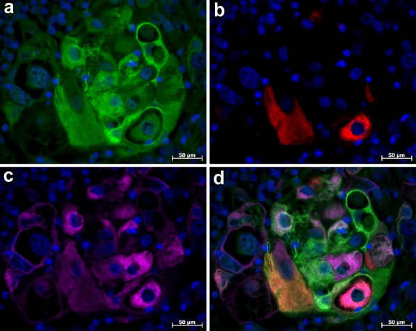
Figure 3. Immunofluorescent triple staining of cytokeratin 5, cytokeratin 10 and cytokeratin 14 in adeno-squamouse carcinoma of human mammary gland.
Immunolabelling was performed without the protein blocking step prior to incubation with primary Abs. (a) Immunolocalization of cytokeratin 14 (Alexa 488, green channel). (b) Immunolocalization of cytokeratins 10 (Cy3, red channel). (c) Immunolocalization of cytokeratin 5 (Alexa 647, pink channel). (d) Composite image. Nuclei are counterstained with DAPI.

