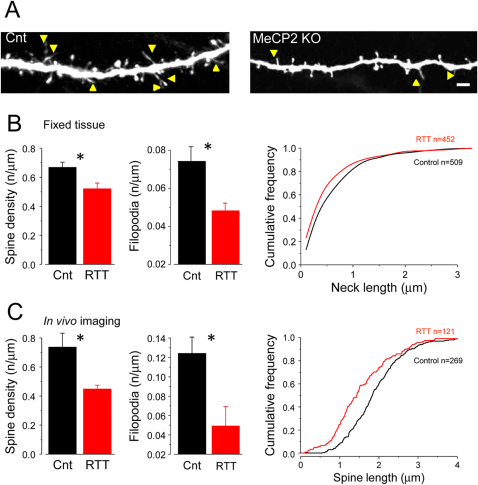
Figure 1. Morphological alterations of dendritic spines in GFP-MeCP2-KO mice.
(A) Confocal micrographs showing GFP-positive dendrites of layer V pyramidal neurons in the S1 cortex of perfused-fixed control (cnt) and GFP-MeCP2-KO mice (RTT). Filopodia-like spines are indicated with arrowheads. Scale bar 2.5 μm. B, C, left and center panels) Both spines and filopodia are less dense in the RTT mice (t-test. B: spines, p<0.03; filopodia, p<0.05; C spines, p = 0.01; filopodia, p = 0.02) when evaluated in fixed tissue (B) or in vivo (C). B, C right panels) Cumulative distributions of spine neck-length (B, fixed tissue) and of spine length (C, in vivo) revealed a general decrement of spine length in the RTT mice (KS-test, B: p<0.01; C: p<0.001). Column plots show the mean and the standard error of the mean.

