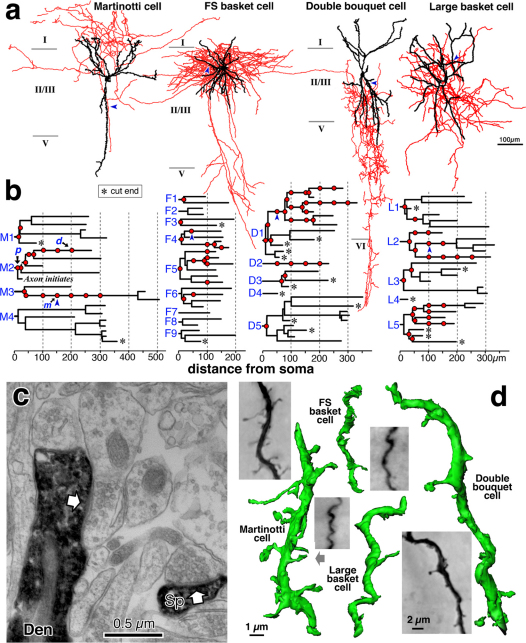
Figure 1. Dendritic structures of neocortical cells representing 4 nonpyramidal neuron subtypes.
(a) Morphology of the neuron. Axons are shown in red, dendrites and somata are shown in black. The MA cell extends its axonal arbors into layer I, whereas the FS cell has a horizontally elongated axonal arborization. The DB cell has a descending narrow axonal arborization, and the LB cell has a large soma and basket terminals. (b) Dendrograms of the same neurons. Red circles indicate segments processed for 3D reconstruction from successive EM sections. Asterisks indicate cut ends of dendrites. Dendritic trees are labeled (M1-M4, F1-F9, D1-D5, L1-L5) and referenced in later figures. Ultrastructure images for segment p, m, d of Martinotti cell are shown in Figure 3 g–i, respectively. (c) Ultrastructure showing dendrite (Den) and spine head (Sp) of the MA cell, which correspond with the dendritic segment indicated by arrow head in (a) and (b). White arrows indicate synapses occurring on those structures. (d) Multi-focus photograph of the dendrites indicated by arrowheads in (a) and (b), along with reconstructed dendrites made from successive EM images of the same dendrite segments. Gray arrow indicates the spine and dendrite shown in (c).

