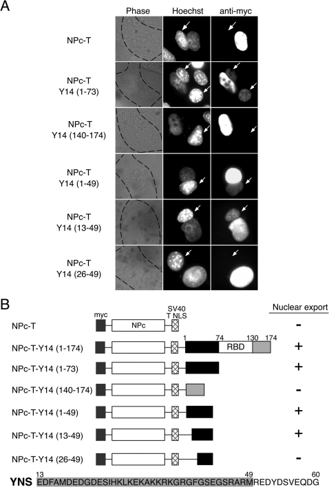
Figure 2. Delineation of the nuclear export signal (NES) of Y14.
(A) Expression vectors encoding various nucleoplasmin core (NPc) fusion proteins were transfected into HeLa cells. After expression of the transfected DNAs, the cells were fused with NIH3T3 cells to form heterokaryons and incubated in media containing 100μg/ml cycloheximide for a period of 2 hrs. The cells were then fixed and stained for immunofluorescence microscopy with a monoclonal antibody 9E10 (anti-myc panel) to detect the fusion proteins, and with Hoechst 33258 (Hoechst panel) that discriminates the human and mouse nuclei within the heterokaryon (the mouse nuclei are shown by arrows). The phase panel shows the phase contrast image of the heterokaryons with the cytoplasmic edge highlighted with black dashed lines. (B) Schematic drawing of the NPc fusion proteins and summary of the heterokaryon results as depicted in (A). The sequences of the minimally defined YNS domain are shown below, and the sequences sufficient for NES activity are highlighted in gray shading.

