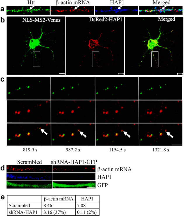
Figure 3. HAP1 is involved in the dendritic targeting of β-actin mRNA.

(a) Co-localization of Htt, β-actin mRNA, and HAP1 in rat cortical neurons (DIV 9). Htt, β-actin mRNA, and HAP1 are shown in green, red, and blue, respectively. The left part of each image is the proximal part of the dendrite. The arrows indicate the co-localization of Htt, β-actin mRNA, and HAP1. Scale bar: 5.0 μm. (b) Co-trafficking of HAP1 with β-actin mRNA in rat cortical neurons (DIV 6). β-actin mRNA is seen in green and HAP1 in red. HAP1 co-localizing with β-actin mRNA is in yellow in the merged panel. Scale bar: 10.0 μm. Part of the boxed area is shown at a high-magnification in (c). (c) Four cropped images from a time-lapse series captured over 1656.5 seconds. The left part of each image is the proximal part of the dendrite. The arrows indicate retrograde movement of one mRNA granule with HAP1 in the neuron. The distance that the granule traveled was 4.03 μm. Scale bar: 5.0 μm. (d) Knockdown of HAP1 decreases β-actin mRNA levels in rat neurons. DIV 4 cortical neurons were infected with lentivirus expressing scrambled shRNA or shRNA-HAP1 and probed at DIV 8 for β-actin mRNA (red), HAP1 (blue), and GFP (green). The left part of each image is the proximal part of the dendrite. Scale bar: 5.0 μm. (e) Quantitative analysis of the intensity of β-actin mRNA and HAP1 normalized to GFP. The percent shown in parentheses indicates extent of knockdown for HAP1, and reduction in signal for β-actin mRNA relative to scrambled.
