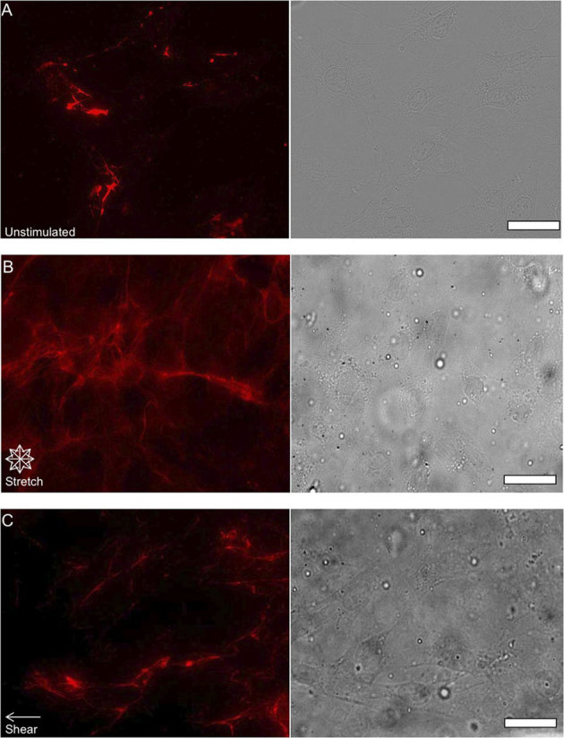
Figure 4. Examining the effects of chemical and mechanical stimulation on FN.

Epifluorescent images of FN (red pseudo color) and phase contrast images of NIH 3T3 fibroblast morphology after inhibition of Rho activity with C3 transferase that were unstimulated (A) after 24 hours of 10% equibiaxial strain (B) and after 24 hours of 6±3 dynes/cm2 of fluid shear stress (C) Cells exposed to mechanical stimulation appear to exhibit an increase in fibril formation as well as forming thicker FN bundles, but at lower densities relative to cells not treated with C3 transferase. (scale bars = 10 μm).
