
Figure 6. Probing cell-substrate interactions of unstimulated cells through high-resolution confocal imaging.
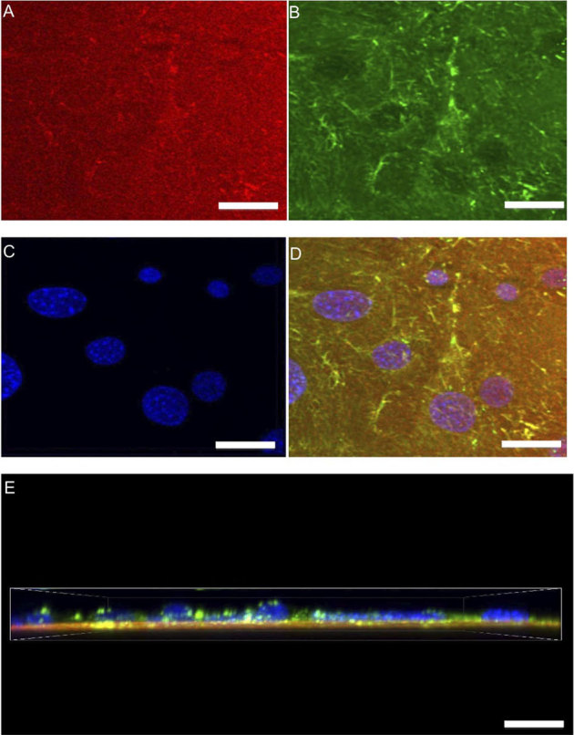
Z-stacks (0.5 μm steps) of unstimulated cells were captured to analyze the labeled substrate FN and its three-dimensional response modified during matrix formation. A top view displays substrate FN (A) cellular fibronectin (B) DAPI (C) and a merged image (D) of all structures. Also a merged side view (E) is presented. Unstimulated cells were observed to have randomly distributed FN fibrils with the substrate FN appearing to not change. (scale bars = 5 μm).