FIGURE 1:
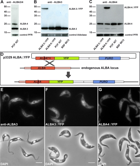
ALBA protein expression and localization in wild-type and ALBA::YFP–expressing cell lines. (A–C) Reactivity of different ALBA antibodies assessed by Western blot in the indicated cell lines (2 μg of total protein extracts per lane). (A) An antibody recognizing both ALBA3 (21 kDa) and ALBA4 (25 kDa) was used on protein extracts from procyclic form 427 (PCF) cells. (B) A specific anti-ALBA3 antibody and (C) an antibody mostly specific for ALBA4 were assessed in cell lines expressing either ALBA3::YFP (46 kDa) or ALBA4::YFP (50 kDa) fusion proteins in PCF wild-type Lister 427 and in bloodstream-form parasites (BSF) of the strain 9013 (Wirtz et al., 1999). (D) Schematic representation of the procedure used for endogenously tagging ALBA proteins. The plasmid p3329, containing a fragment of the ALBA gene upstream of the YFP sequence and a puromycin-resistance cassette, is linearized (red line) within the ALBA sequence before transfection. This allows homologous recombination with one of the endogenous ALBA copies to create YFP-tagged ALBA. (E) Cultured procyclic cells were analyzed by IFA using the anti-ALBA3 antibody (top) and counterstained with DAPI (white, bottom) merged with the phase contrast image. Cells of the Lister 427 strain expressing (F) endogenously ALBA3::YFP or (G) ALBA4::YFP were fixed with PFA and counterstained with DAPI (white, bottom). Scale bar, 5 μm.

