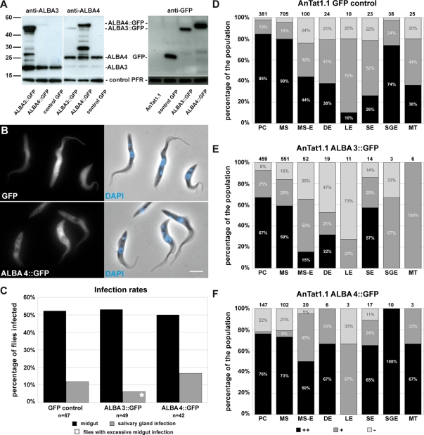
FIGURE 5:
Overexpression of ALBA3::GFP or ALBA4::GFP and consequences for fly infection. (A) Western blots with 2 μg of total protein samples of AnTat1.1 WT cells carrying a GFP gene (control GFP) or an extra copy of either ALBA3::GFP or ALBA4::GFP. Blots were incubated with a specific anti-ALBA3 antibody to detect the endogenous ALBA3 (expected molecular weight 21 kDa) and the ALBA3::GFP (46 kDa) protein (left), with a specific anti-ALBA4 antibody to detect the endogenous ALBA4 (25 kDa) and the ALBA4::GFP (50 kDa) protein (middle), or an anti-GFP antibody to reveal the control GFP protein (right) and the ALBA::GFP fusion proteins. A weak cross-reactivity of the anti-ALBA4 antibody with ALBA3 is visible. Anti-PFR antibody was used as a control. (B) Procyclic cells of the strain AnTat1.1 in culture expressing either GFP or exogenous ALBA4::GFP were fixed in PFA and counterstained with DAPI (left, direct fluorescence; right, phase contrast image and DNA in blue; scale bar, 5 μm). (C) Percentage of flies infected (in the midgut or the salivary glands) when challenged with AnTat1.1 cells expressing GFP control (sum of seven infection experiments; 214 flies, 67 dissected), ALBA3::GFP (sum of six infection experiments; 155 flies, 49 dissected), or ALBA4::GFP (sum of three infection experiments; 143 flies, 42 dissected) (n = total number of dissected flies). Infections with the ALBA3::GFP cell line in the salivary gland were only observed in flies with a highly infected midgut (star). (D–F) Percentage of bright (black), positive fluorescent (dark gray), and negative cells (light gray) in the populations of each stage of AnTat1.1 parasites expressing GFP alone (D), ALBA3::GFP (E), or ALBA4::GFP (F) during the course of infection. Analysis was performed on live trypanosomes directly upon fly dissection as described in the text; the number of counted cells is indicated on top of the bars. To reach the total numbers of parasites, data from all independent infection experiments were grouped for analysis as indicated in C. Cells are shown in the order of appearance during the infection cycle in the tsetse fly: procyclic (PC), mesocyclic trypomastigote (MS), parasites in transition from mesocyclic trypomastigote to epimastigote (MS-E), and asymmetrically dividing epimastigote (DE) into a long (LE) and a short epimastigote (SE); in the salivary glands, salivary gland epimastigote (SGE) and metacyclic (MT).

