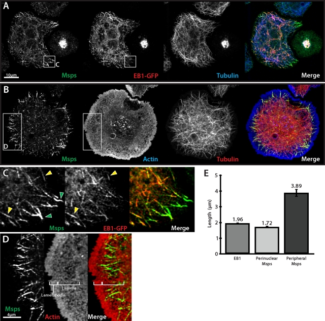
FIGURE 1:
Msps localizes to both microtubule plus ends and to the lattice of peripheral microtubules. (A) Interphase Drosophila S2 cell transfected with EB1::EB1-GFP (middle) and immunostained for Msps (left) and α-tubulin (right). (B) S2 cell immunostained for Msps (left), actin (middle), and α-tubulin (right). (C) Inset from A, Msps colocalizes with EB1 and peripheral microtubules that are EB1 negative. (D) Inset from B, Msps lattice accumulations (left) are coincident with actin-rich lamella (right). (E) Graph of microtubule decoration length for immunostained EB1 and interior-localized and peripheral Msps. Error bars, 95% CI.

