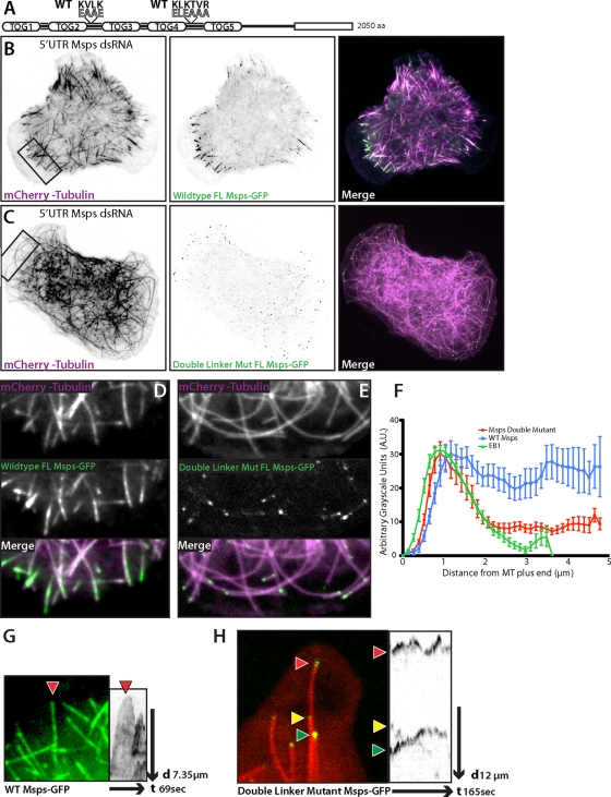
FIGURE 6:
Mutation to the linker regions of Msps abrogates interaction with the lattice of peripheral microtubules. (A) Cartoon schematic of full-length double-mutant Msps (Double Mut Msps) that contains the linker2 KVLK charge reversal and linker4 charge reversal. (B) Interphase S2 cell depleted of endogenous Msps using 5′ UTR dsRNA, expressing wild-type Msps-GFP (B) (Supplemental Movie S6) or Double Mut Msps-GFP (C). (D–E) Inset of B (left) and C (right) with Msps-GFP (top left) or Msps Double Linker Mut (top right), mCherry-Tubulin (middle), and the merge (bottom). (F) Line scans across plus ends of S2 cells expressing EB1::EB1-GFP, full-length wild-type Msps-GFP, or full-length double-mutant Msps-GFP (Double Mut Msps). Line scans for Msps are of peripheral microtubules at equal exposures and time frames; error bars, SD. (G) (Supplemental Movie S7) Peripheral microtubule decorated with wild-type Msps-GFP and the associated kymograph. (H) (Supplemental Movie S8) Peripheral microtubule end with Double Mut Msps-GFP and kymograph.

