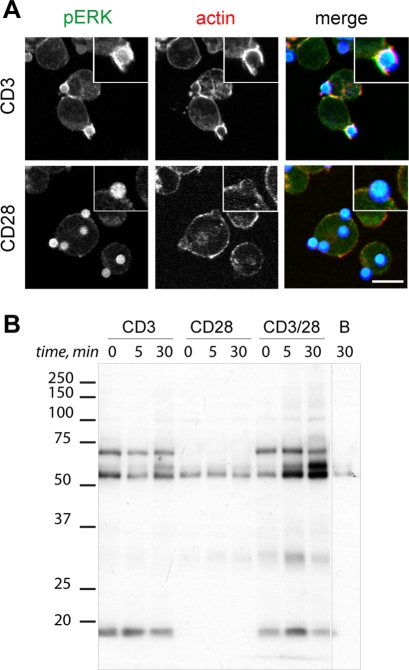
FIGURE 1:
Isolation of TCR immune complexes using anti-CD3/CD28–coated beads. (A) Jurkat T cells were stimulated with sheep anti–mouse IgG Dynabeads coated with human anti-CD3 or anti-CD28 (10 min). Immunostaining is shown for activation (pERK) and actin dynamics markers (phalloidin–rhodamine). Insets, zoomed area. Bar, 10 μm. (B) Jurkat T cells (107 cells/ml) serum starved in DMEM, 0.5% BSA (2 h), were incubated with anti-CD3, anti-CD28, or anti-CD3/CD28 on ice, followed by Dynabeads (37°C) for indicated times. Time 0 shows cells incubated with antibody and Dynabeads on ice. Associated protein complexes were resolved by SDS–PAGE and tyrosine-phosphorylated proteins developed in WB. Left, molecular weight markers. Control immunoprecipitation was performed with Dynabeads alone (lane B).

