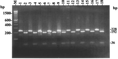
FIG. 3.
Confirmation of the mycobacterial origin of PCR amplicons (228 bp) based on NarI restriction patterns (192 and 36 bp). Shown are gel patterns for M. chelonae reference strain (lane 1 and 2) and for NTM field isolates M-JY1 (lanes 3 and 4), M-JY2 (lanes 5 and 6), M-JY3 (lanes 7 and 8), M-JY4 (lanes 9 and 10), M-JY5 (lanes 11 and 12), M-JY6 (lanes 13 and 14), M-JY7 (lanes 15 and 16), and M-JY8 (lanes 17 and 18); lane M, 100-bp DNA size marker (Invitrogen). For each isolate, the two lanes represent the original amplicon (8 μl) and its NarI restriction digestion product, respectively.

