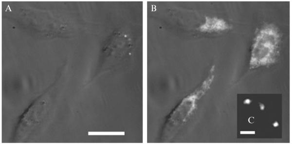
Figure 2.2. Photoactivation of the azido DCDHF fluorogen in live mammalian cells.
(A) Three Chinese Hamster Ovary cells incubated with azido DCDHF fluorogen are dark before activation. (B) The fluorophore lights up in the cells after activation with a 10-s flash of diffuse, low-irradiance (0.4 W cm−1) 407-nm light. The white-light transmission image is merged with the fluorescence images, excited at 594 nm (~1 kW cm−1). Scalebar: 20 μm. (C) Single molecules of the activated fluorophore in a cell under higher magnification. Scalebar: 800 nm. (Adapted with permission from Lord et al., 2008. Copyright 2008 American Chemical Society.)

