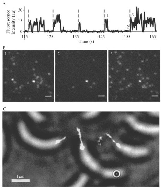
Figure 2.4. Photoswitching behavior of and super-resolution imaging using the Cy3–Cy5 covalent dimer.
(A) Representative single-molecule fluorescence time trace of 5-labeled bovine serum albumin showing reactivation cycles 12–16, denoted by the dashed lines. (B) Fluorescence images at times 1, 2, and 3 corresponding to the times labeled in panel (A). Scale bar, 1 μm. (C) Super-resolution fluorescence image of C. crescentus stalks with 30 nm resolution superimposed on a white-light image of the cells. The C. crescentus cells were incubated in 4 μM of Cy3–Cy5 NHS ester for 1 h and then washed five times before imaging to remove free fluorophores. The data were acquired over 2048 100-ms imaging frames with 633 nm excitation at 400 W cm−2. After initial imaging and photobleaching of the Cy3–Cy5 dimers, the molecules were reactivated every 10 s for 0.1 s with 532-nm light at 10 W cm−2. (Adapted and reproduced with permission from Conley et al., 2008. Copyright 2008 American Chemical Society.)

