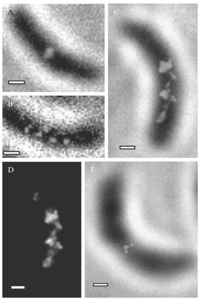
Figure 2.6. Super-resolution images of MreB in live C. crescentus cells.
(A–B) Images taken using standard PALM (C–D) images taken using time-lapse imaging to obtain higher labeling density using. (A) Image of MreB forming a midplane ring in a predivisional cell. (B) Banded MreB structure in a stalk cell. (C) Quasi-helical MreB structure at 40 nm resolution observed using time-lapse PALM. (D) Structure in panel (C) displayed without white-light image in order to highlight the continuity of the structure. (E) Time-lapse PALM image of MreB midplane ring in a predivisional cell. (Adapted and reproduced with permission from Biteen et al., 2008. Copyright 2008 Nature Publishing Group.)

