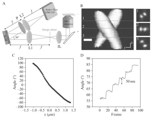
Figure 2.7. Localization of 200-nm fluorescent beads using the DH-PALM microscope.
(A) Schematic illustrating the collection path in a DH-PALM setup, IL is imaging lens, L1 and L2 are 150 mm focal length achromat lenses, P is a linear polarizer, and SLM is a phase-only spatial light modulator. (B) Three-dimensional representation of the experimentally observed DH-PSF (created with VolumeJ (Abrámoff and Viergever, 2002) with slices taken at z positions of approximately (1) −450 nm, (2) 0 nm, and (3) 500 nm where 0 is taken to be the designed focal plane of the microscope. (C) Calibration of angle between the two lobes as a function of the distance between the objective surface and the bead with 0 being the position when the lobes are horizontal with respect to one another. (D) Plot of angle between the lobes versus frame for a fluorescent bead as the objective is scanned through 50 nm steps showing clear steps in the angle with a low standard deviation in each step.

