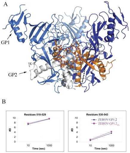
Figure 2. The fusion loop of ZEBOV-GP1,2.
(A) Cartoon representation of the fusion loop of GP2 (shown in ball-and-stick representation with carbon atoms colored orange). GP1 subunits of the 3-fold related protomers are shown in different shades of blue. One of the GP2 subunits is shown in orange, and the other two are shown in grey. The crystallographically disordered loop that is cleaved by cathepsin L/B is shown as a dotted line. (B) Deuteration plots of the residues in the fusion loop in ZEBOV-GP1,2 (shown in dark blue) and ZEBOV-GP1,2CL (shown in magenta).

