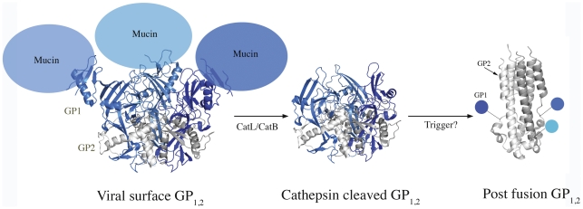
Figure 5. Changing structures of ZEBOV-GP1,2.
Cartoon representations of GP1,2 in its viral surface form (PDB: 3CSY), putative receptor-binding form and post-fusion form (PDB: 2EBO). GP1s and GP2s are colored in different shades of blue and grey, respectively. The mucin-like domains were deleted from ZEBOV-GP1,2 for crystallization and have been modeled here as not-to-scale balloons. It is currently unclear if GP1 remains attached to GP2 during the conformational changes that lead to fusion. The transmembrane regions at the bottom of GP1,2 are not illustrated.

