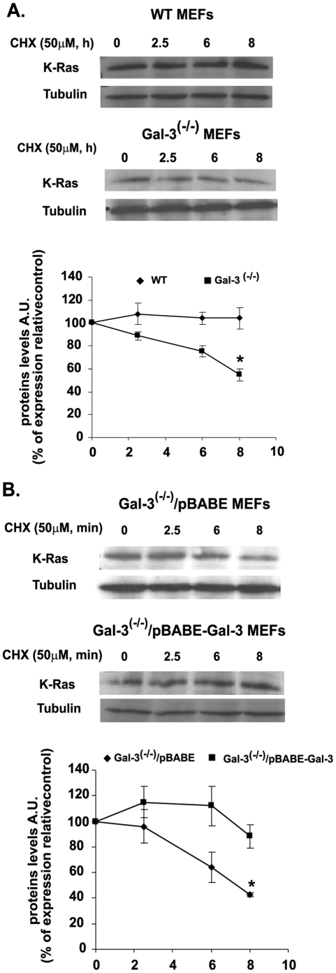
Figure 3. Gal-3 attenuates K-Ras protein degradation.
A. Attenuation of K-Ras degradation in wt and Gal-3-/- MEFs. Cells of the wt and Gal-3-/- MEFs were grown (2×10⁵ cells per 6-cm plate) in the presence of 10% FCS, which was replaced 24 h later by 0.5% FCS. Cells were treated with cycloheximide (CHX; 50 µg/ml) and then harvested at zero time (control) and at 2.5, 6, and 8 h after CHX treatment. K-Ras in the lysates was quantified by SDS–PAGE followed by immunoblotting with anti-K-Ras Abs. β-Tubulin served as a loading control. Upper panel: typical immunoblots visualized by ECL. Lower panel: expression levels of K-Ras, normalized to β-tubulin as a percentage of the expression at zero time (means ± SEM, n = 3 *p<0.05). B. Attenuation of K-Ras degradation in Gal-3-/- vector-infected and Gal-3-/- pBABE-infected MEFs. Stable cell lines (see Methods) were treated with CHX and then harvested and immunoblotted as in A. Upper panel: typical immunoblots visualized by ECL. Lower panel: expression levels of K-Ras, normalized to β-tubulin as a percentage of the expression at zero time (means ± SEM, n = 3 *p<0.05). MEFs, mouse embryonic fibroblasts; wt, wild type.

